
Objectes multimèdia amb l’etiqueta: Centres docents
Resultats de la cerca

Cuerpos hinchados
Accés obert
17 d’oct. 2022
Conferència a càrrec de l'arquitecte Smiljan Radic´per Projectes I-II T, assignatura coordinada per Pere Joan Ravetllat, Departament de Projectes Arquitectònics ETSAB | UPC.
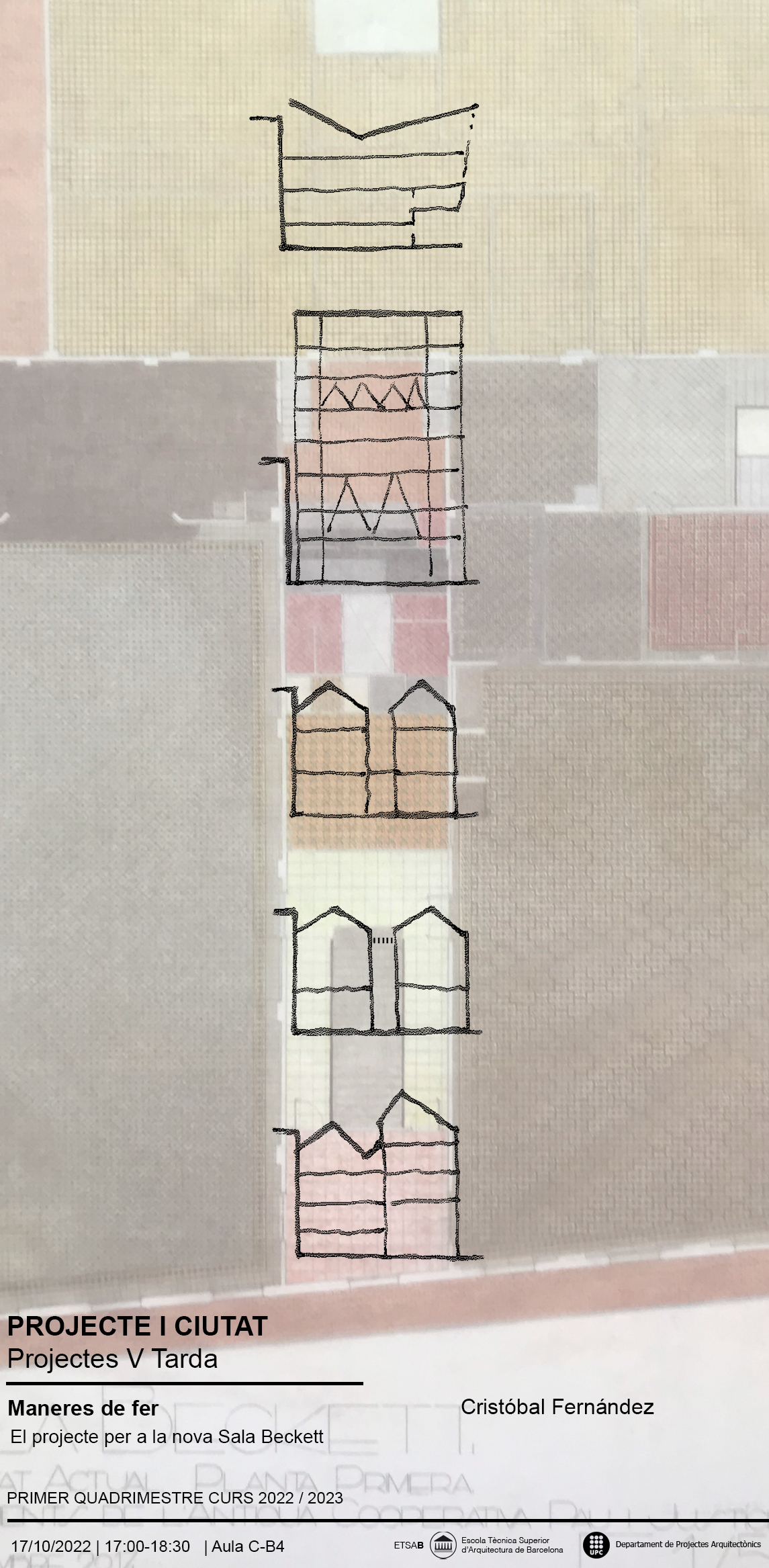
Maneres de fer. El projecte per a la nova Sala Beckett
Accés obert
17 d’oct. 2022
Conferència de Cristóbal Fernández, professor del Departament de Projectes Arquitectònics ETSAB | UPC per Projecte i Ciutat, Projectes V T, assignatura coordinada per Félix Solaguren Beascoa i Berta Bardí (DPA ETSAB | UPC).
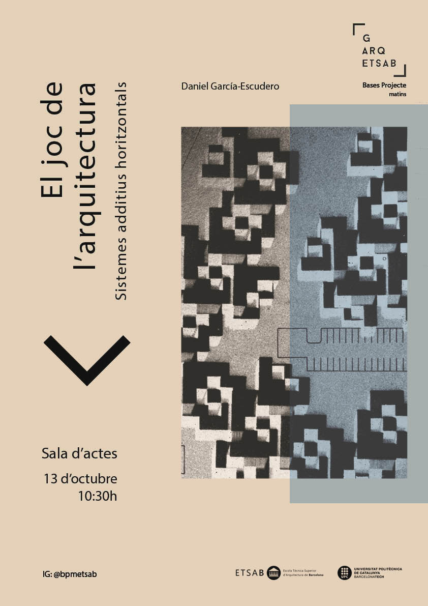
El joc de l'Arquitectura: sistemes additius horitzontals
Accés obert
13 d’oct. 2022
Conferència a càrrec de Daniel García-Escudero (Projectes Arquitectònics ETSAB | UPC), professor coordinador de l'assignatura Bases Projecte I-II [m].
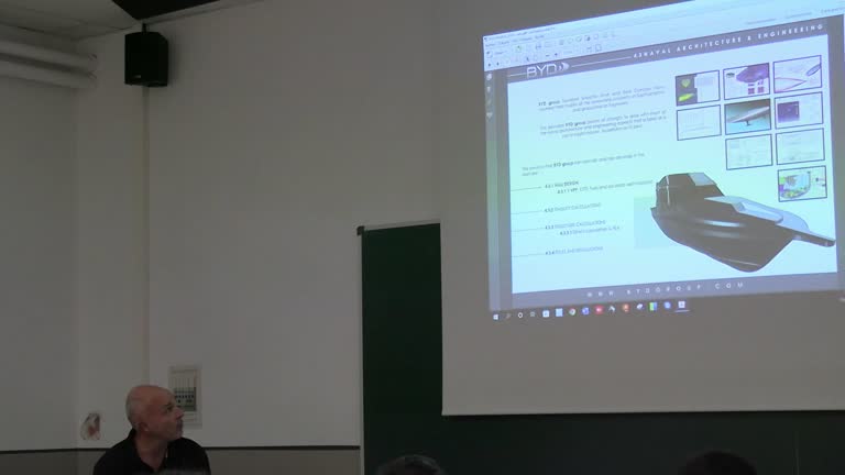
Trabajos de una oficina de diseño, Ingeniería y Arquitectura Naval: BYD Group
Accés obert
11 d’oct. 2022
Presentació del treball de l'oficina fundada per Raúl Gonzalo i Sebastián Simó de Barcelona Yacht Design, Naval Architecture and Engineering. Introducció a càrrec del degà Luis Fernandez Cotelo degà del Colegio de Ingenieros Navales y Oceánicos, delegació de Catalunya, i professor de l'FNB. Xerrada adreçada especialment als estudiants del Màster en Enginyeria Naval i Oceànica de l'FNB.
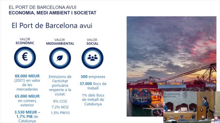
Inauguració del curs acadèmic 2022-2023
Accés obert
11 d’oct. 2022
Obertura de l'acte pel degà de l'FNB Sr. Agustí Martín Mallofré. Presentació de la Memòria del curs acadèmic 2021/22, a càrrec del Sr. Ramon Grau Mur, Secretari Acadèmic de l'FNB. Lliçó inaugural a càrrec de l'honorable Sr. Damià Calvet i Valera, President del Port de Barcelona. Lliurament regals commemmoratius als millors expedients de fase inicial. Cloenda a càrrec del vicerector de Transferència, Innovació i Emprenedoria de la UPC, Sr. Climent Molins Borrell.
Inauguració del curs 2022-2023
Accés obert
5 d’oct. 2022
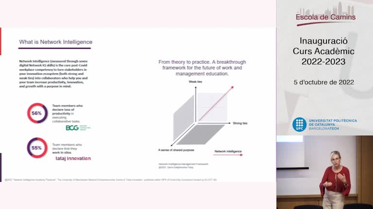
El dimecres 5 d’octubre, a les 12 hores a la Sala d’Actes Jose Antonio Torroja de l’Escola de Camins va tenir lloc la inauguració del curs acadèmic 2022-2023.
La conferència inaugural: "Network Intelligence and deeptech entrepreneurial talent" va anar a càrrec de la Dra. Daria Tataj, professora Honoraria de Alliance Manchester Business School, creadora de Network Intelligence (nIQ) methodology, Fundadora & CEO de Tataj Innovation, i autora de “Innovation and Entrepreneurship: A Growth Model for Europe beyond the Crisis”.
En el decurs de l'acte es va fer un reconeixement a diversos membres de l’Escola de Camins i es van lliurar els premis als estudiants amb millors expedients acadèmics.
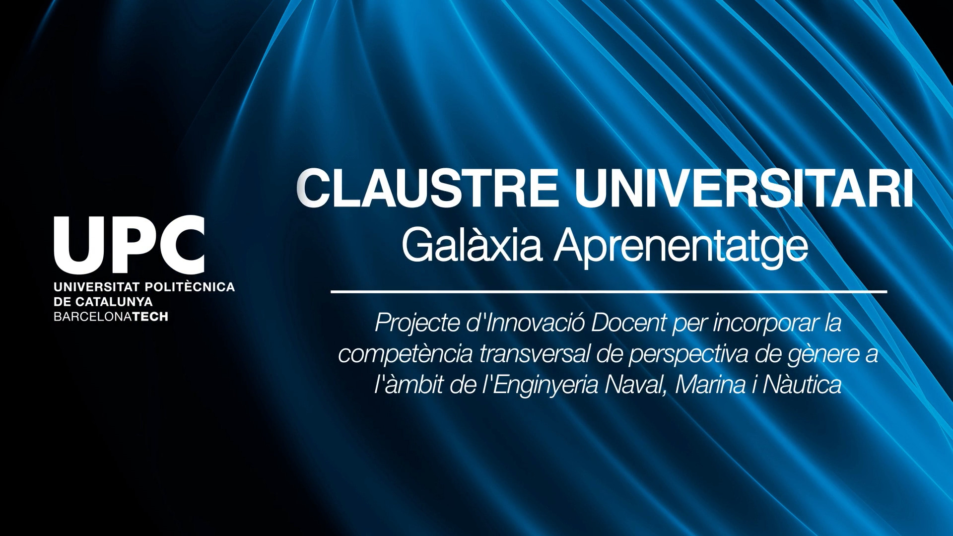
FNB: Incorporant la competència transversal de perspectiva de gènere als estudis
Accés obert
3 d’oct. 2022
Clàudia Barahona, Xavier Martínez, Santiago Ordàs i Marcel·la Castells, professors de la Facultat de Nàutica de Barcelona (FNB), expliquen com han incorporat la la competència transversal de perspectiva de gènere als estudis de l'àmbit de l'Enginyeria Naval, Marina i Nàutica.
La reflexió i el debat sobre l’experiència d’aprenentatge a la Universitat i les línies estratègiques per a un nou model docent centren el Claustre Universitari de la UPC, els dies 4 i 7 d’octubre.
La reflexió i el debat sobre l’experiència d’aprenentatge a la Universitat i les línies estratègiques per a un nou model docent centren el Claustre Universitari de la UPC, els dies 4 i 7 d’octubre.
Marc Alier: Reflexions
Accés obert
29 de set. 2022
Marc Alier Forment, professor de la Facultat d'Informàtica de Barcelona (FIB) i del Departament d'Enginyeria de Serveis i Sistemes d'Informació, i sotsdirector de l'Institut de Ciències de l'Educació (ICE) de la UPC, explica una experiència d'innovació docent.
La reflexió i el debat sobre l’experiència d’aprenentatge a la Universitat i les línies estratègiques per a un nou model docent centren el Claustre Universitari de la UPC, els dies 4 i 7 d’octubre.
La reflexió i el debat sobre l’experiència d’aprenentatge a la Universitat i les línies estratègiques per a un nou model docent centren el Claustre Universitari de la UPC, els dies 4 i 7 d’octubre.
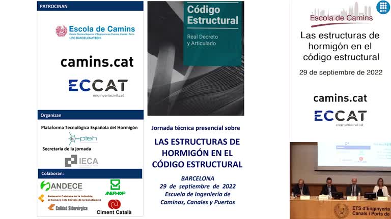
Inauguración de la Jornada Técnica sobre las Estructuras de Hormigón en el Código Estructural.
Accés obert
29 de set. 2022
La Plataforma Tecnológica Española del Hormigón y el Instituto Español del Cemento y sus Aplicaciones, con el apoyo de la Escuela de Caminos, el Colegio de Ingeniería Técnica de Obras Públicas y de Ingeniería Civil y el Colegio de Ingeniería de Caminos, Canales y Puertos organizó una jornada técnica sobre el Código Estructural.
Jordi Guàrdia: Mind the gap!
Accés obert
29 de set. 2022
Jordi Guàrdia Rubies, professor de l'Escola Politècnica Superior d'Enginyeria de Vilanova i la Geltrú (EPSEVG) i del Departament de Matemàtiques de la UPC, explica una experiència d'innovació docent.
La reflexió i el debat sobre l’experiència d’aprenentatge a la Universitat i les línies estratègiques per a un nou model docent centren el Claustre Universitari de la UPC, els dies 4 i 7 d’octubre.
La reflexió i el debat sobre l’experiència d’aprenentatge a la Universitat i les línies estratègiques per a un nou model docent centren el Claustre Universitari de la UPC, els dies 4 i 7 d’octubre.
